


Single Cell Optical Manipulations
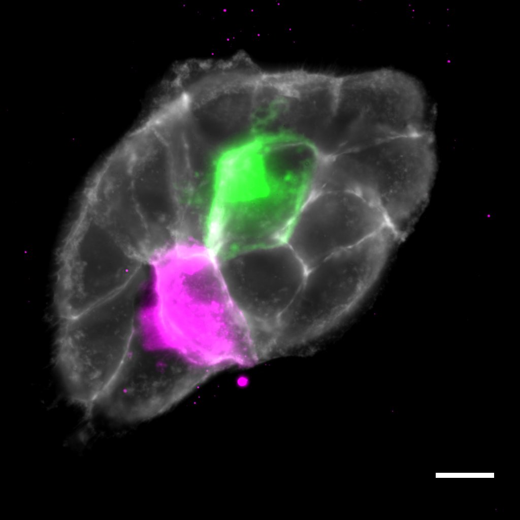
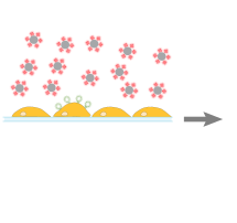
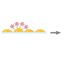
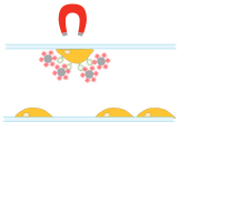
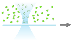
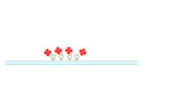
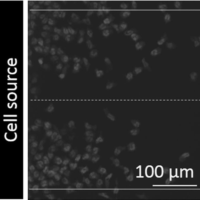
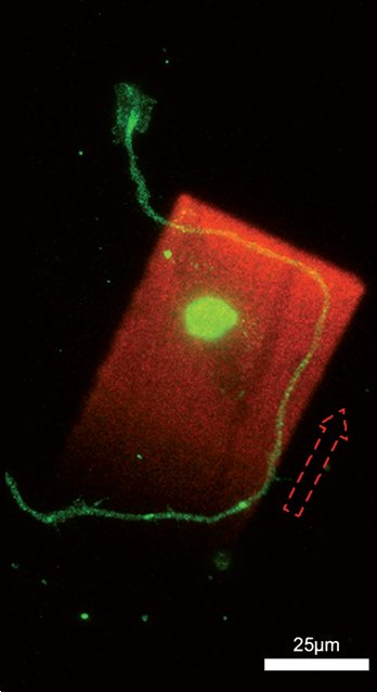
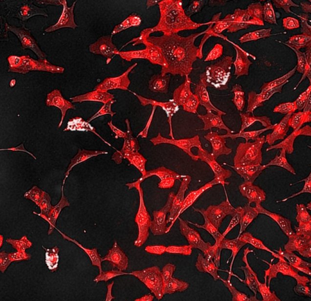
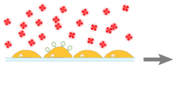
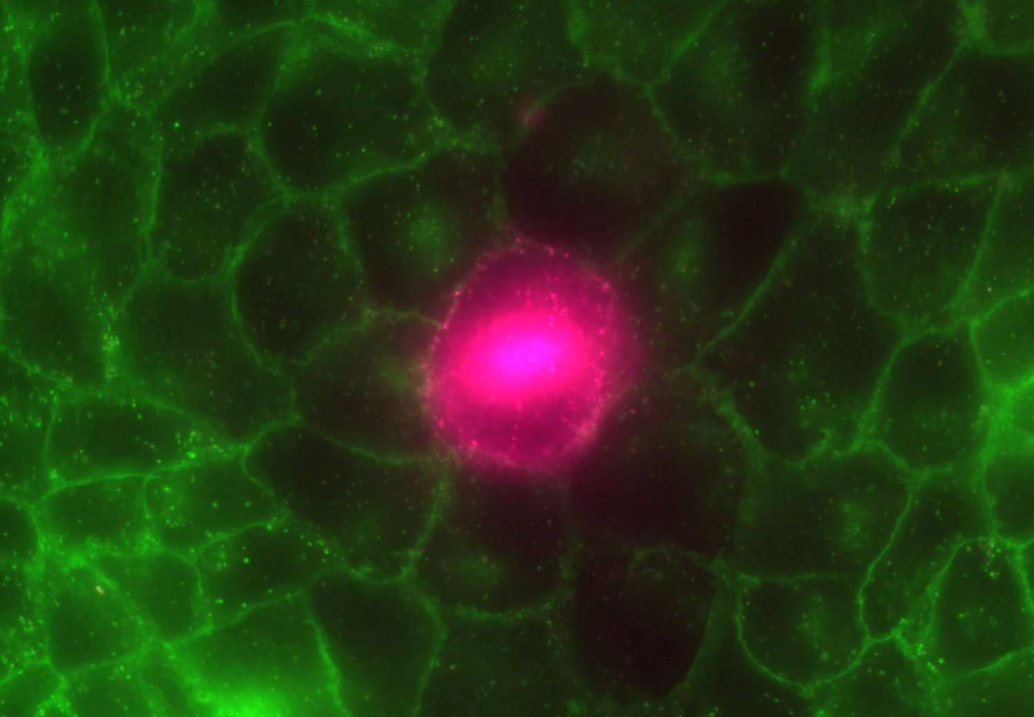
CLaP takes advantage of the lasers of a confocal microscope to bind biotin molecules to the plasma membrane of individually selected cells. Streptavidin of different colors are used to label cells or streptavidin-coated ferromagnetic beads and a simple magnet can be used to capture only illuminated cells with high purity. This novel technology allows to explore many biological processes of interest in the fields of axon guidance, cell migration, cancer biology, immunology and many more.